
Christian Coachman, Marcelo A Calamita and Eric Van Dooren present a realistic and practical treatment plan.

Part one: background, case objectives and treatment plan
The ‘new world’ of aesthetic dentistry has created a great burden for the practising clinician. Media-centred advertising would have one believe that anyone and everyone can buy the ‘perfect’ smile. Although most of us recognise that this is not the case, the global hype has created a number of negative consequences:
- Patients present with unrealistic expectations
- Many dentists are misguided into thinking that they should be able to meet or exceed patients’ aesthetic expectations in all cases. Many of these clinicians feel inadequate if they cannot achieve the results that they see in dental journals, magazines and online resources
- Some cosmetic dentists are focusing more on patient ‘wants’ rather than ‘needs’ and are not availing themselves of a comprehensive approach to treatment.
This case was chosen because it is does not exemplify ‘ideal’ treatment. It is not a treatment plan that the present-day cosmetic dentist would necessarily want to implement, or one that the treating clinician would consider as a first, or even second choice of therapy. However, it is a realistic and practical plan, considering the amount of bone loss present in the aesthetic zone. It is also novel in its design, as well as minimally invasive and simple in concept, and the outcome was aesthetically pleasing.
Case presentation
The treating clinicians were prosthodontist, periodontist and implant surgeon, Eric Van Dooren; orthodontist Ralph Lemmens DDS and dental laboratory and smile designer, Christian Coachman, DDS, CDT.
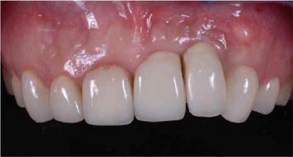
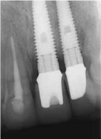
A 42-year-old woman presented with an aesthetic problem associated with a failing implant restoration. The previous treatment, necessitated by trauma resulting from a car accident, was performed seven years prior to the initial examination. Extensive bone grafting was performed at that time, in order to restore the anterior ridge defect. Two implants had been placed in the areas of the missing teeth, UL1 and UL2.
Treatment planning considerations
- The patient was apprehensive about treatment because of negative past dental experiences and expressed an interest in a non-invasive, nonsurgical approach. She qualified her wishes by stating that if surgery was absolutely unavoidable to achieve a successful aesthetic result, it should be minimal and limited to a single procedure
- The patient expressed some concerns about her smile, especially regarding the midline, inclination of the incisal plane, and lack of symmetry in the length and form of her anterior teeth
- The patient wanted to focus on the failing implants and did not want any of her other teeth to be treated. On the other hand, she understood that in order to improve tooth proportions and to create more aesthetic harmony and balance, additional teeth might require treatment.
Summary of concerns
- Even if the patient consents to surgical soft and hard tissue augmentation, are there any surgical techniques that would predictably and fully restore the anterior ridge to its original state? Is it advisable to propose surgery, even with qualifications, and possibly create unrealistic expectations?
- Considering that this patient is hesitant to proceed with any surgical treatment, if a decision is made to replace the existing crowns and to try to restore harmony and balance to the face and smile, how can this be accomplished with minimally invasive procedures?
- Recognising that we may be limited in the amount of vertical ridge augmentation that can be achieved, how will we be able to compensate for this restoratively to meet or exceed the patient’s treatment expectations?
Proposed treatment plan
Goals/objectives of treatment:
- Accomplish a fixed method of rehabilitation without involving non-restored teeth
- Achieve optimal aesthetics.
Phase one: data gathering and digital planning:
- Intraoral and full-face digital photos to be captured in order to establish:
- Proper incisal edge position
- Appropriate midline position and angulation
- Ideal tooth proportions and gingival contours.
Photographic documentation is transferred to the computer for digital planning. The first step would be to relate the full-face smile photo to the horizontal reference line. The most common reference line used is the interpupillary line, but the nasal or commissural lines can also be used. The midline is then projected onto the horizontal line and digital planning initiated. The treatment plan is evaluated, modified and related to the digital smile design (DSD).
Phase two: transfer of digital wax-up to the master cast:
- The digital plan is transferred to the master cast
- Tooth form is copied from the digital forms
- White aesthetics are optimised
- Pink aesthetics are optimised
- The 3D defect around the implants is filled with pink wax, which allows us to evaluate the vertical and horizontal components of the ridge deficiency.
Phase three: fabrication of intraoral mock-up and provisional partial denture:
- The digital project or intraoral smile design is evaluated
- Old restorations are removed, tooth locations and form evaluated. Anterior provisional partial denture is fabricated
- DSD is evaluated and minor changes in form made by applying flowable composite. The size of the anterior ridge defect is visualised by both the patient and the dentist
- Decisions are made as to whether and to what extent the anterior defect could be managed and how this will affect the definitive restoration. Much of the decision-making is based on careful 3D evaluation of the soft tissue contours, with special focus on the papilla heights and volume
- Aesthetic and functional evaluation are performed to establish incisal edge position, soft tissue contours, altered midline inclination, lip support and tooth form, which should provide overall facial balance and harmony
- Patient approval is obtained
- The treatment plan is finalised.
Phase four: establishing the definitive treatment plan:
- Any final changes to the treatment plan are made, taking into consideration patient preferences. Final adjustments are also made to maximise soft tissue harmony, gain optimal soft tissue contours (compensating for the ridge deficiency), and optimise selection of prosthetic materials
- The definitive digital and intraoral concept is translated into the definitive treatment plan.
Phase five: orthodontic therapy:
Orthodontic treatment is initiated in the mandibular arch to realign the mandibular anterior teeth and to intrude the incisors to help reduce the existing vertical overlap.
Phase six: surgical treatment:
- Surgical therapy is performed, including clinical crown lengthening to attain adequate biologic width in the maxillary anterior segment
- Connective tissue grafting is performed where indicated to augment the anterior ridge
- Postoperative care is provided, and healing allowed to occur.
Phase seven: definitive restorations:
- Minimally invasive definitive tooth preparation is performed
- Final impressions are taken, and the master cast is fabricated
- Definitive restorations are fabricated
- All restorations are tried in
- The definitive restorations are cemented.

