Case of the month: a radiology report due to problems in patient anatomy

Figure 1: Reconstructed panoramic radiograph

Images provided

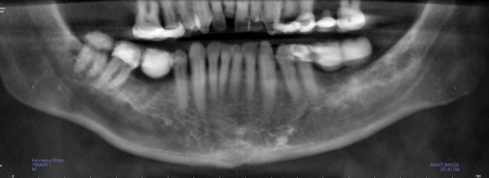
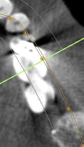
Cone beam CT images in the bone window. Axial, coronal and sagittal planes.

Clinical info

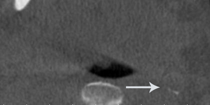
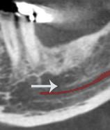
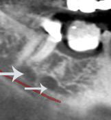
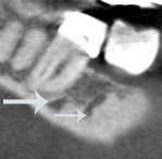
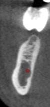
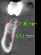
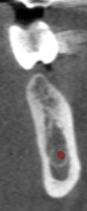
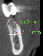
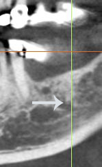
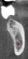
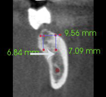
Implant analysis requested. Relevant history – please see comments and special requests. Client notes – the primary areas of concern are those sites involved in future implant placement, the LR5 and LL5 and LL6. In the LL5 site I have noticed a well corticated area close to the buccal plate, roughly 5mm from the crest of the ridge. Similarly in the LR5 site there is a round radiopaque area 9mm from the crest of the ridge towards the lingual plate. My concern is that these are arteries that I would like to avoid.

Diagnostic objectives

  1. Implant planned
  2. Rule out pathology.

Findings

Mandible: localised areas of increased vascularity were observed superior to the mandibular canal in the mandibular right and left molar region.

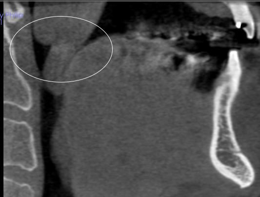
Air space: there appears to be an ovoid area of increased soft tissue density superior and posterior to the uvula and the soft palate. In addition, the pharyngeal tonsils appear to be enlarged, especially the right side.

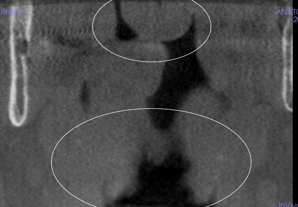
Other findings: curvilinear areas of increased density were noted within the lower neck in areas anatomically associated with the carotid arteries; these areas appear to be consistent with calcification the carotid artery.

Dental findings: no abnormalities detected.

Radiographic impression

Air space: clinical evaluation of the soft tissues of the oral pharynx is suggested. Physician referral for more thorough evaluation of the potential soft tissue mass superior and posterior to the soft palate is recommended to rule out neoplasia.

Other findings: the small areas of increased density noted within the lower neck are anatomically associated with the carotid arteries and are consistent with calcification of the carotid arteries. Vascular calcifications have been associated with an increased risk of cardiovascular disease and stroke. Review of patient’s medical history for increased risk factors such as high blood pressure, elevated cholesterol, stress, and smoking is suggested. Physician referral is suggested with findings of elevated risk factors or if patient is not currently under the care of a physician.

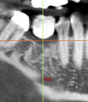
Implant measurements: have been provided for requested sites; please review the comments associated with each of the cross-sections.

For more information visit ct-dent.co.uk.

Untitled-2
Figure 2: Curvilinear areas of increased density were noted within the lower neck in areas anatomically associated with the carotid arteries; these areas appear to be consistent with calcification the carotid artery


Figure 3: There appears to be an ovoid area of increased soft tissue density superior and posterior to the uvula and the soft palate. In addition, the pharyngeal tonsils appear to be enlarged
Figure 4: There appears to be an ovoid area of increased soft tissue density superior and posterior to the uvula and the soft palate. In addition, the pharyngeal tonsils appear to be enlarged
Figure 5: There appears to be an ovoid area of increased soft tissue density superior and posterior to the uvula and the soft palate. In addition, the pharyngeal tonsils appear to be enlarged
Figure 3: There appears to be an ovoid area of increased soft tissue density superior and posterior to the uvula and the soft palate. In addition, the pharyngeal tonsils appear to be enlarged
Figure 6: There appears to be a localised increase in vascularity in the mandibular left first molar region (arrows)
Figure 8: There appears to be a localised increase in vascularity in the mandibular left first molar region (arrows)
Figure 7: There appears to be a localised increase in vascularity in the mandibular left first molar region (arrows)
Figure 8: Localised areas of vascularity were observed in the mandibular right first and second molar region
Figure 9: Localised areas of vascularity were observed in the mandibular right first and second molar region
Figure 10: Localised areas of vascularity were observed in the mandibular right first and second molar region
Figure 11: Mandibular right first molar region along the green line
Figure 12
Figure 12
Figure 13
Figure 13
Figure 14: Mandibular left first molar region along the green line. The arrow indicates the superior edge of the area of increased vascularity previously described
Figure 14: Mandibular left first molar region along the green line
Figure 16
Figure 15
Figure 16
Figure 16: The arrow indicates the superior edge of the area of increased vascularity previously described
Figure 17: Mandibular left second molar region along the green line
Figure 18: The arrow indicates the superior edge of the area of increased vascularity previously described
Figure 19
Figure 19
Figure 20
Figure 20


Favorite
Get the most out of your membership by subscribing to Dentistry CPD
  • Access 600+ hours of verified CPD courses
  • Includes all GDC recommended topics
  • Powerful CPD tracking tools included
Register for webinar
Share
Add to calendar