Cone Beam CT images in the bone window. Axial, coronal and sagittal planes.
Clinical information
Implant analysis requested. Relevant history – confirm absence of pathology (including in apical area UL2, UL3, where on peri-apical films there has been a historic radiolucency). Potential implant treatment planning for either removable implant supported overdenture or fixed bridgework.
Diagnostic objectives
Implant planned
Sinus evaluation
Rule out pathology.
Findings
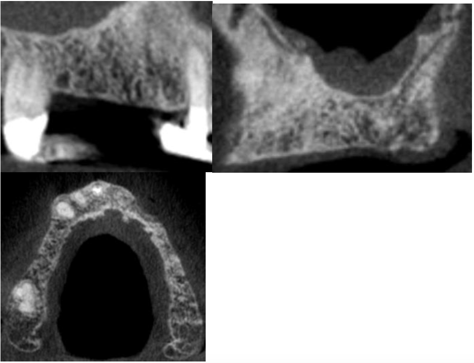
Maxilla: cross-sections of the anterior maxilla have been provided below illustrating an irregular area of low-density in the alveolar process to the left of the midline.
Sinuses: mild to moderate increases in the thickness and density of the tissues lining the right and left maxillary sinuses were noted.
Nasal cavity: a mild deviation of the nasal septum was noted.
Other findings: no abnormalities detected.
Dental findings: no abnormalities detected.
Radiographic impression
Maxilla: please see the detailed descriptions associated with the cross-sections illustrating the maxilla.
Sinuses: the radiographic findings appear to be consistent with a mild mucositis/sinusitis of the right and left maxillary sinuses. Review of the patient’s clinical history for chronic allergy or sinusitis is suggested; physician referral for more thorough evaluation is suggested if merited by clinical findings and symptoms.
Nasal cavity: deviation of the nasal septum is a common incidental radiographic finding and does not require treatment or referral unless the patient exhibits difficulty breathing through the nose.
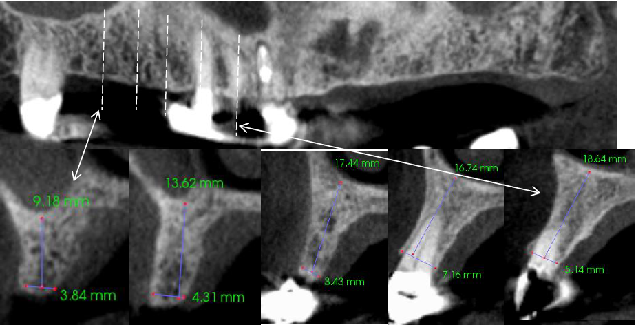
Implant measurements:have been provided for various areas the maxilla and may not correspond to the final sites selected for implant placement but provide an indication of the bone available in various areas.
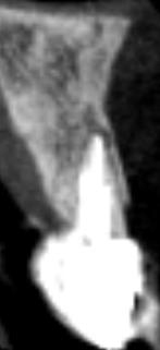
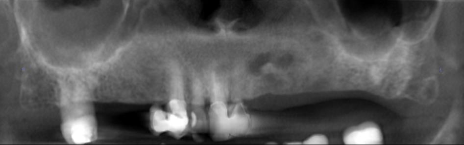
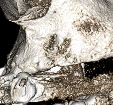
Mild to moderate increases in the thickness and density of the tissues lining the right and left maxillary sinuses were noted. A mild deviation the nasal septum was notedBuccal 3D rendering of the radiolucent area in the maxillary left lateral incisor-canine regionLingual/palatalThe maxillary left lateral incisor-canine region exhibits a large irregular area of radiolucency/low density extending through the alveolar process labial and linguallyCross-sections of the maxillary anterior region with the cross-sections oriented with the long axis of the incisive canal and foramen. The irregular radiolucency observed to the left of the midline appears to exhibit a potential communication/connection with the incisive canal and foramen (single arrows). The large arrow illustrates a potential vascular canal on the lingual surface of the alveolar process, which in other cross-sections communicates with the irregular area of radiolucency. Radiologist comment: the irregular area of radiolucency observed in the maxillary left lateral canine region may potentially represent a surgical defect created at the time the teeth were removed; however, the potential communication of the area with the incisive canal and foramen is highly suggestive of a large vascular anomaly in this area. Advanced medical imaging is suggested prior to any planned surgical procedures in the area to rule out a vascular lesion. The double headed arrow indicates a small dilation of the incisive canal to the right of the midline suggestive of a small, early cyst of the incisive canalA well circumscribed periapical radiolucency was noted surrounding the apex of the maxillary right central incisor. Clinical evaluation of the tooth is suggestedCross-sections through the right maxilla; note the prominence of the vascular canals visible in the cross-sectionsCross-sections through the left maxillaRadiologist comment: the entire maxilla exhibits a highly increased visibility of vascular structures not typically observable. The general radiographic appearance is suggestive of a highly vascular maxillary bone as surgical procedures in the area may be accompanied by increased bleeding. Review of the patient’s medical history for possible systemic manifestations of hemangiomas in other parts of the body suggested to rule out a systemic disorder of vascularity

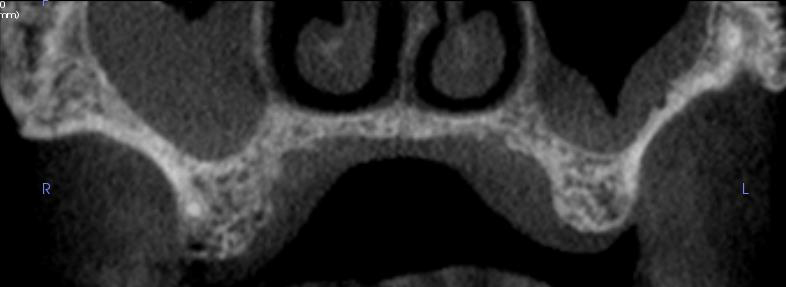


![Cross-sections of the maxillary anterior region with the cross-sections oriented with the long axis of the incisive canal and foramen. The irregular radiolucency observed to the left of the midline appears to exhibit a potential communication/connection with the incisive canal and foramen [single arrows]. The large arrow illustrates a potential vascular canal on the lingual surface of the alveolar process which in other cross-sections communicates with the irregular area of radiolucency. Radiologist comment: the irregular area of radiolucency observed in the maxillary left lateral canine region may potentially represent a surgical defect created at the time the teeth were removed; however, the potential communication of the area with the incisive canal and foramen is highly suggestive of a large vascular anomaly in this area. Advanced medical imaging is suggested prior to any planned surgical procedures in the area to rule out a vascular lesion. The double headed arrow indicates a small dilation of the incisive canal to the right of the midline suggestive of a small, early cyst of the incisive canal](https://www.dentistry.co.uk/app/uploads/2016/09/Screen-Shot-2016-09-13-at-10.52.22.png)