CT Dent presents a case where 3D imaging was used to identify the extent of hidden incisal trauma.
This dentoalveolar trauma case resulted from a fall from a bike, requiring an assessment of the upper anterior teeth to rule out fractures.
Radiographic impression
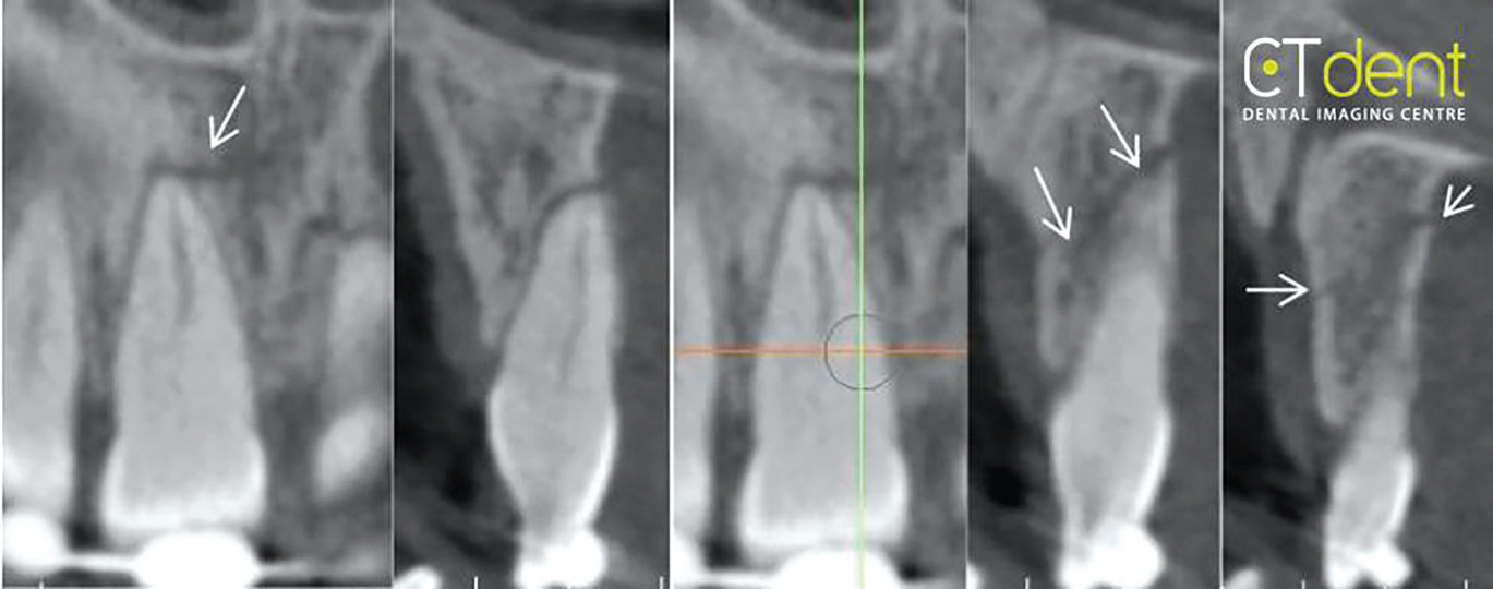
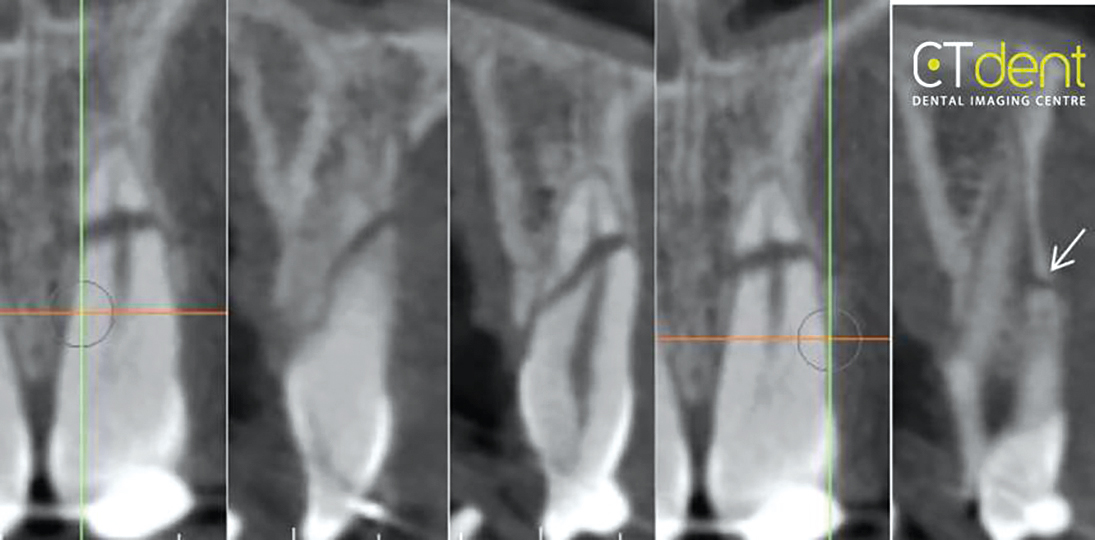
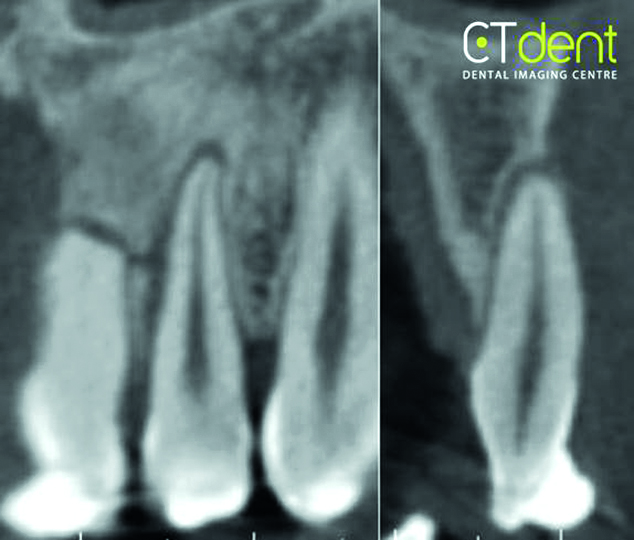
Fracture of the maxillary anterior alveolar process extending from the mesial surface of the right central incisor to the interproximal space between the maxillary left central and lateral incisors. Fracture through the apex of the left central incisor also seen. Periodontal ligament (PDL) spaces associated with the right central incisor and left lateral incisor are consistent with partial traumatic avulsion.
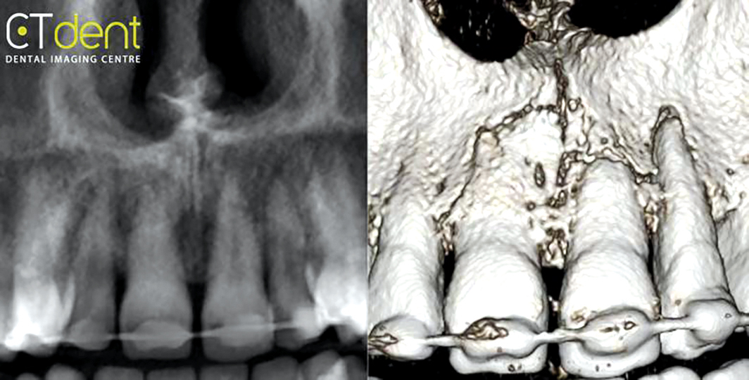
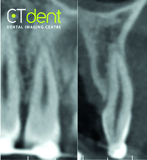
Figure 1: Reconstructed panoramic radiographFigure 2: Maxillary right lateral incisor – the tooth appears to be normalFigure 3: Maxillary right central incisor region – the PDL space is widened and there appears to be a line of fracture (arrow) adjacent to the apex of the root. A line of fracture through the alveolar process was observed adjacent to the mesial edge of the right central incisorFigure 4: Maxillary left central incisor – the line of fracture extends through the apical third of the left central incisor and exits through the labial surface of the alveolar process adjacent to the distal surface of the central incisorFigure 5: Maxillary left lateral incisor region – the PDL space surrounding the lateral incisor is widened and consistent with partial traumatic avulsion
For more information about CT Dent, visit ct-dent.co.uk.