vVARDIS presents two case reports demonstrating guided enamel regeneration as an effective clinical solution for early caries management.
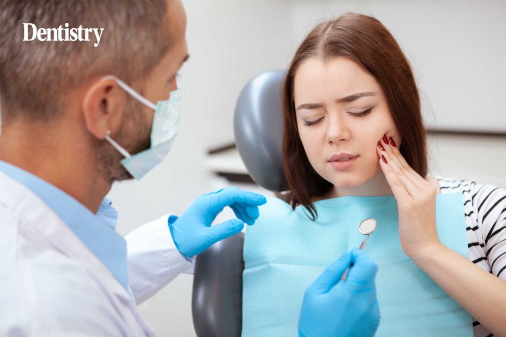
Traditional management of early carious lesions often involves a passive ‘watch-and-wait’ approach until cavitation occurs, at which point invasive treatment is required. However, biomimetic treatments now offer clinicians the ability to intervene earlier, preserving natural tooth structure and improving patient outcomes.
CURODONT REPAIR, based on self-assembling peptide technology (P11-4), creates a 3D matrix within the lesion body, guiding hydroxyapatite formation using calcium and phosphate from the patients’ saliva. This approach promotes remineralisation without drilling, discomfort, or anaesthesia, making it ideal for both aesthetic and structural enamel remineralisation.
The following cases demonstrate how CURODONT REPAIR can successfully arrest and reverse early lesions, offering patients a biologically sound alternative to traditional restorations.
Case 1: post-orthodontic white spot lesion management
Presentation
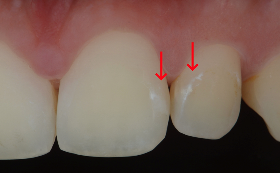
A 25-year-old male patient presented with a concern from white discolouration on the maxillary anterior teeth following fixed orthodontic treatment.
Examination
White spot lesions were visible on the labial surfaces of teeth 21 and 22 (Figure 1). The lesions evident both wet and dry.
Diagnosis
Initial enamel caries (ICDAS Score 2) on teeth 21 and 22, attributed to inadequate plaque control during orthodontic treatment.
Treatment
One in-surgery application of CURODONT REPAIR was performed. The patient was also prescribed CURODONT PROTECT for at-home use twice weekly at bedtime.
Outcome
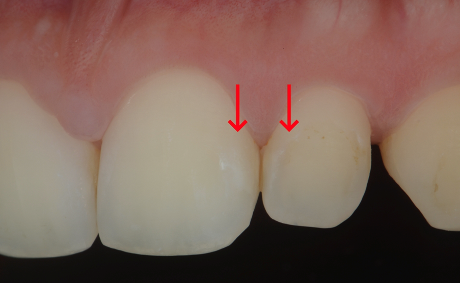
At the 16-month follow-up, the lesions showed a reduction in both size and opacity, indicating effective caries regression and aesthetic improvement (Figure 2). The patient reported high satisfaction.
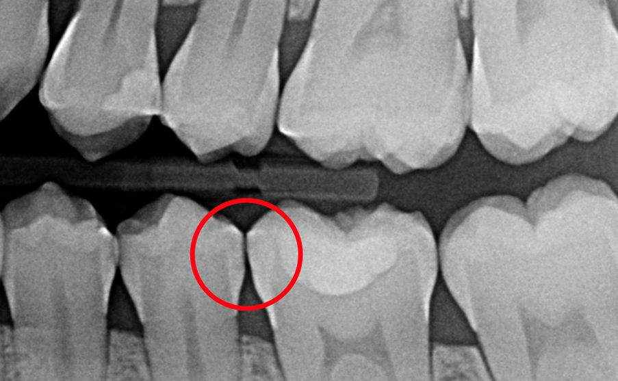
Case 2: early interproximal lesion in a high caries-risk patient
Presentation
Incidental finding during routine recall of a 28-year-old female patient with high caries risk.
Examination
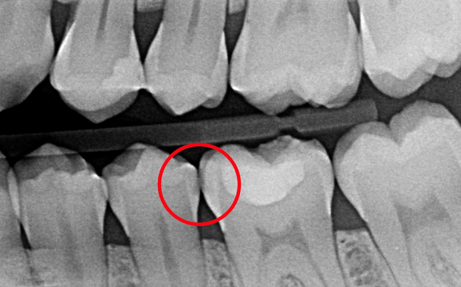
Bitewing radiograph revealed a radiolucent lesion on the distal surface of tooth 35 and mesial surface of tooth 36 (Figure 3).
Diagnosis
35: Initial distal enamel lesion (E2), 36: Moderate mesial dentine lesion.
Treatment
CURODONT REPAIR was applied to teeth 35 and 36. The patient was prescribed CURODONT PROTECT twice weekly.
Outcome
At the seven-month follow-up, the lesion showed signs of arrest and reduced radiolucency (Figure 4), indicating enamel regeneration.
Clinical takeaway
These cases demonstrate how CURODONT REPAIR can effectively treat both early caries white-spot lesions and interproximal lesions in patients. By avoiding drilling, local anaesthetic, and discomfort, this approach reduces patient anxiety and enhances trust. More importantly, it supports true biological healing through guided enamel regeneration, preserving enamel rather than replacing it.
Cases provided by Dr Giovanni Sammarco, Cariologia Clinica, Quintessence Publishing Italia, 2025.
For more information, please contact your local Henry Schein representative.
This article is sponsored by vVARDIS.

